- Tooth Extractions or Exodontia
- Transalveolar Extraction
- Surgical Endodontic Procedure
- Pre-Prosthetic Surgery
- Exposure of Unerrupted Teeth
- Biopsy and Cysts Removal
Dentoalveolar Surgery
Dentoalveolar surgery refers to all surgical procedures involved with the teeth and associated structures i.e. bone and soft tissues. Dr. S.M. Balaji has extensive surgical experience and performs the surgical procedure(s) quickly and with less surgical trauma.
Tooth extractions or exodontia
Teeth may need to be removed for many reasons including severe decay, advanced gum disease, infection or as part of an orthodontic treatment plan.
-
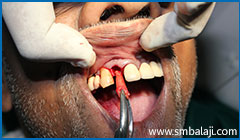
Exodontia or tooth extraction
-

X-ray taken before extraction of upper right third molar tooth
-
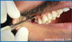
During extraction- Accessing the tooth from the cheek sid
-
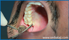
During extraction- accessing the tooth from the tongue side
-

During extraction- Grasping the tooth with forceps
-
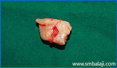
The extracted upper right third molar
Sometimes a tooth may fail to emerge into proper alignment and remains “entrapped” in the gum tissue and jaw bone. Such tooth is referred to as impacted. Impactions may occur due to various reasons. For example, the jaw may be too small and there may be insufficient space for the teeth to erupt. Teeth may also become twisted, tilted, or displaced as they try to emerge, resulting in impacted teeth.
The most common impacted teeth are wisdom teeth or third molars. Impacted teeth may result in swelling, pain and infection of surrounding gum tissue. Also, impacted teeth may cause permanent damage to nearby teeth or lead to the formation of cysts or tumors that can destroy sections of the jaw. Therefore, it is often recommend that impacted teeth be promptly removed.
CASE I
-
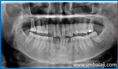
X-ray showing two impacted lower canine teeth
-

X-ray taken before extraction of upper right third molar tooth
-
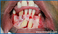
Surgical exposure of impacted teeth
-

Immediately after surgical removal of impacted lower canine teeth
-
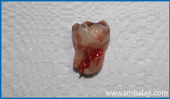
Impacted wisdom tooth surgically removed atraumatically
CASE II
-
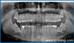
X-ray showing 6 impacted teeth- upper right and left third molars and lower right and left third and second molars
-
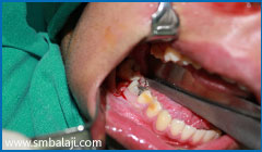
Surgical exposure of lower right impacted teeth
-
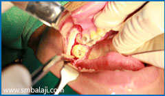
Surgical exposure of lower left impacted teeth
-
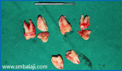
All impacted teeth removed
CASE III
-

X-ray showing teeth impacted in the lower right jaw region
-
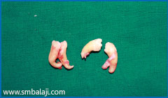
Impacted teeth surgically removed
CASE IV
-
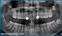
X-ray showing impacted upper right and left third molar teeth
-
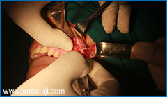
Left tooth surgically exposed
-
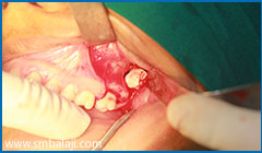
Left tooth surgically exposed
-
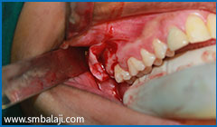
Right tooth surgically exposed
-
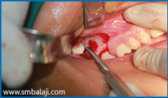
Right tooth surgically exposed
-
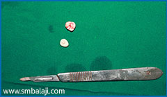
Impacted teeth surgically removed
The most common of these is a surgical apicoectomy, which is usually performed on a root canal treated tooth that has become infected or painful. An apicoectomy is a procedure in which the gum tissue near the tooth is opened to see the underlying bone and infected tissue is removed. The very end of the root tip is also removed and the root canal filling is sealed.
-
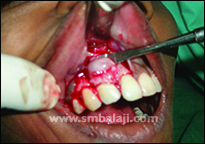
During apicoectomy- infection at root tip exposed
-

Apicoectomy done
-
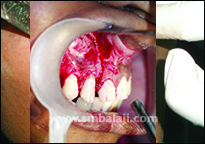
Apicoectomy done
-
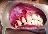
Immediately after apicoectomy
-
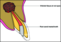
Severe infection at the root tip
-
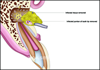
Infection and root tip removed in apicoectomy procedure
Sometimes surgical modifications of the jaw bone and soft tissues are required to enable the placement of a well-fitting, comfortable and aesthetic dental prosthesis such as dentures or dental implants. This preparation is referred to as pre-prosthetic surgery.
It is important that the bony ridge and soft tissues be of proper size and shape before placing the prosthesis. To achieve this, one or more of the following procedures might be needed
- Bone smoothing and reshaping (alveoloplasty)- irregular or sharp bone ridges are smoothened off
- Bone grafting – this is required to “fill in” the bony defects. When there is missing teeth, jaw bone tends to shrink over time. There may not be sufficient bone to place dental implants or prosthesis. Or bone defects may occur due to gum disease. Bone grafting is a procedure to rebuild bone in the bone deficient areas of the jaw. For this, bone may be harvested from other parts of the jaw, patient’s rib or bone substitutes may be used.
- Removal of excess bone (exostoses)- large and irregular chunks of excess bone may need to be removed to allow denture to fit properly.
- Adding, removing or reshaping the soft tissues- the soft tissues inside the mouth may require to be surgically corrected to have a well-fitting, stable denture. Excess soft tissue may require to be removed or some tissues may need to be repositioned to ensure a successful prosthesis. Soft tissue grafting may sometimes be required.
CASE I
-
-image-of-the-lower-jaw-showing-mandibular-tori-(exostoses).jpg)
3D Cone Beam CT (CBCT) image of the lower jaw showing mandibular tori (exostoses)
-
-image-of-the-lower-jaw-showing-mandibular-tori-(exostoses)-.jpg)
3D Cone Beam CT (CBCT) image of the lower jaw showing mandibular tori (exostoses)
-

Clinical photograph showing the tori
-
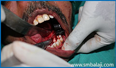
Tori surgically excised from the lower left jaw region
-
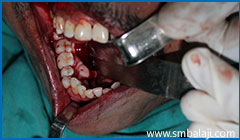
Tori surgically excised from the lower right jaw region
-
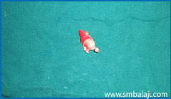
A portion of the excised bone excess
CASE II
-

Severely insufficient bone in missing teeth region of upper jaw and slanting bite
-
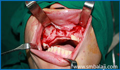
Deficient region surgically exposed
-
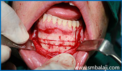
Mandibular segmental subapical osteotomy - excess bone removal from left lower jaw to correct slanting bite
-
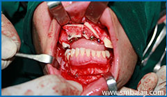
Implant placement in upper jaw after bone grafting with excess bone from lower jaw
CASE III
-
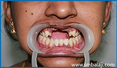
Missing upper front teeth with insufficient bone and altered alignment of upper and lower jaw
-
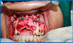
Le Fort I maxillary osteotomy and upper jaw advancement
-
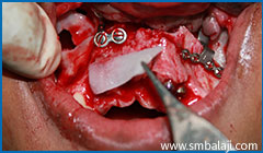
rhBMP-2 placed to stimulate new bone formation and implants placed
-
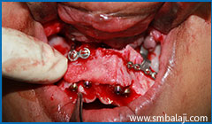
rhBMP-2 will stimulate new bone formation reinforcing the bone deficient region
-
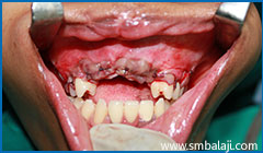
Upper jaw adequately advanced and brought into more favourable position in relation to lower jaw
-
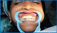
After completeion of prosthetic rehabilitation with ceramic crowns and correct teeth alignment
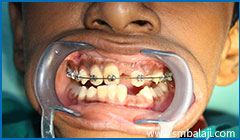
Exposure and bonding of unerupted teeth for orthodontic purposes – some teeth do not erupt normally and remain buried in the jaw bone. Unlike impacted wisdom teeth which are often removed, these teeth (e.g. cuspid teeth in upper jaw) are important for good dental form and function. Such “hidden” teeth may need to be surgically “uncovered” by a procedure called operculectomy and an orthodontic appliance needs to be fixed to them that will gradually bring the tooth into normal position
CASE I
-
3D Cone Beam CT (CBCT) image of the lower jaw showing mandibular tori (exostoses)
-

3D Cone Beam CT (CBCT) image of the lower jaw showing mandibular tori (exostoses)
-
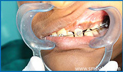
Clinical photograph showing the tori
-
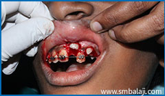
Tori surgically excised from the lower left jaw region
Biopsy and removal of simple cysts and other lesions of the mouth – Some lesions occurring in the mouth and maxillofacial region required to be examined for any disease. A tissue sample is taken from the lesion and sent for histopathological examination. This procedure is called a biopsy. Some simple lesions and infections including cysts have to be removed.
CASE I
-
-showing-extensive-cyst-in-lower-jaw-middle-region-and-upper-left-jaw.jpg)
3D Cone Beam CT image (CBCT) showing extensive cyst in lower jaw middle region and upper left jaw
-

Digital X-ray showing cyst lesions
-
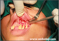
Bony lesion surgically exposed in the upper jaw
-
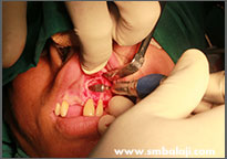
Cyst lesion surgically removed in upper jaw
-
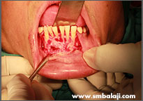
Tori surgically excised from the lower right jaw region
-
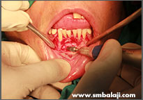
Cyst lesion surgically removed in lower jaw
-
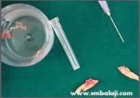
Excised lesions and extracted infected teeth
-
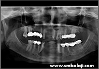
X-ray following surgery showing good bone healing
CASE II
-
-image-showing-impacted-tooth-and-cyst-in-lower-jaw-right-side.jpg)
3D Cone Beam CT (CBCT) image showing impacted tooth and cyst in lower jaw right side
-

X-ray showing right lower impacted tooth with cyst lesion and other multiple impacted teeth
-

Surgical exposure of impacted tooth
-
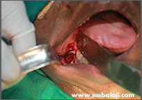
Impacted tooth surgically removed
-
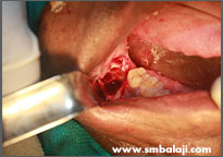
Cyst lesion excised
-
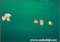
Removed impacted teeth
-
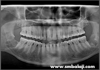
X-ray showing good bone healing after surgery
CASE III
-
-image-showing-impacted-lower-right-wisdom-tooth.jpg)
3D Cone Beam CT (CBCT) image showing impacted lower right wisdom tooth, affected second molar and large cyst lesion in right lower jaw
-
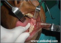
Clinical exposure of the diseased portion of the right lower jaw
-
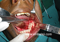
Surgical exposure of the impacted tooth and cyst
-
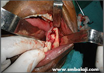
Impacted teeth and cyst excised surgically
-
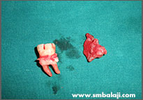
Removed infected tooth and cyst
-
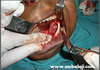
After cyst excision and surgical removal of affected teeth
-

Digital X-ray taken immediately after surgery
-

X-ray showing very good bone healing just three months following surgery
CASE IV
-

X-ray showing cyst lesion in upper jaw right side
-
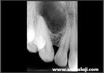
Radiovisuograph showing cyst lesion
-
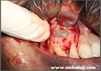
Surgical exposure of cyst
-
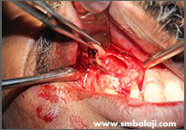
Cyst lesion surgically excised
-
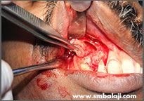
Cyst lesion surgically excised
-
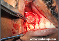
Cyst lesion surgically excised
-
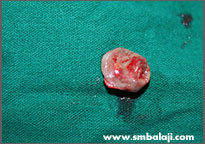
The excised cyst pathology
-
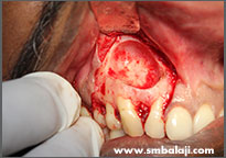
Bone defect cleaned after cyst removal

